
Sutures of the Skull
How many Sutures are in the skull?
The human skull consists of 20 sutures, which means that there are 40 bones in the skull fused together. At birth, a human has a total of 45 skull bones. A skull suture is also called cranial suture. The skull is a vital structure as it encloses one of the most important organs in the body, the brain.
The infant’s skull differs from that of the adult skull because it consists of six separate skull bones namely the frontal bone, occipital bone, two temporal bones and parietal bones. (1, 2, 3)
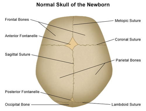
Picture 1: An image showing a typical/normal newborn’s skull.
Photo Source: stanfordchildrens.org
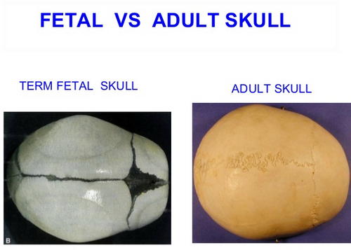
Picture 2: A comparison image between a fetal skull and an adult skull.
Photo Source: image.slidesharecdn.com
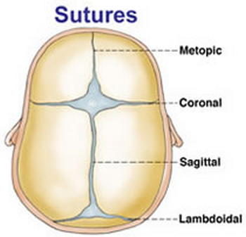
Picture 3: The different types of skull sutures.
Image Source: neurosurgery.ufl.edu
What are the sutures of the skull made up of?
A suture is a line or a junction of articulation between the skull’s adjacent bones. A suture is a fibrous joint type, which is fused together by Sharpey’s fibres. It is the Sharpey’s fibres that allow a bit of flexibility.
The skull looks a lot like a single large bone, but in reality, it consists of several bones connected together. These bones are frontal bones, parietal bones, and occipital bone. (2, 3)
What are the sutures on the human skull?
There are various sutures on the skull. What are the four sutures of the skull? The primary sutures of the skull are coronal, sagittal, squamosal, and lambdoid.
Coronal sutures
It is located between the frontal bone and two parietal bones. Its function is to join frontal bone to the parietal bones. It covers the frontal fontanelle of an infant.
Sagittal suture
It is located in between the two parietal bones. It fuses two parietal bones together.
Squamous suture/squamoparietal suture
It is located between the squamous area of the temporal bone and parietal bone, connecting the parietal bones to the temporal bone.
Lambdoid suture
It is the area between the occipital bone and two parietal bones. It connects the parietal bone to the occipital bone. (4, 5, 6, 7)
Other sutures on the skull include:
Metopic suture
it runs midline of the frontal bone and fuses between three months and nine months of age.
Frontal sphenoid
It usually closes at the age of three months old.
Sphenoparietal suture
It is a tiny suture that separates the parietal bone from the sphenoid bone.
Sphenosquamous suture
It separates the squamous portion of the temporal bone from the sphenoid bone.
Parietomastoid suture
It separates the parietal bone from the temporal bone’s mastoid process.
Occipitomastoid suture
It is the suture that separates the mastoid process from the occipital bone. (6, 7, 8)
The spaces between the skull bones of an infant where the sutures intersect are called fontanelles. They are covered with a strong membrane, which protects the brain and the underlying soft tissues. The fontanelles are divided into two:
- Anterior fontanelle/Soft spot – It is the junction where the parietal and frontal bones meet. It remains soft until the child reaches 18 months to two years old.
- Posterior fontanelle – It is the junction between the occipital bone and parietal bones. It closes during the first few months of the baby’s life. (2, 4, 5)
Functions of sutures in the skull
Sutures function as an expansion joint, which allows the bones to move during the birth process. It allows the head of the newborn to deform so that it can fit through the narrow birth canal. As the brain grows, the skull evenly expands leading to a symmetrical shape of the head.
An abnormal shape of the head happens if any of the sutures prematurely closed as they will hinder the growth in a particular area of the skull. In adult, sutures in the skull enable impact force to be evenly distributed around the skull. (5, 6, 7)
When do skull sutures close completely?
Ideally, the sutures on the skull are completely closed around the age of 2. The sutures will ossify or harden to fuse the bones of the skull together. (6, 7)
What skull bone is not joined by a suture?
The only non-sutured bones in the skull are the mandible and zygomatic bones. (8)
Medical conditions related to sutures of the skull
In the first two years of life, the brain of the child grows rapidly causing the sutures to stretch, which sends a signal to the suture to form new bones. The suture enlarges thereby creating enough space for the growing brain.
This type of suture remains open until the child reaches adulthood. However, when the suture closes earlier than expected, it leads to an abnormal shape of the skull or medically known as craniosynostosis.
A misshapen skull is a result of the brains continuous growth even if the sutures are already closed. It affects not just the shape of the skull but also the brain’s health. There is a possibility of blindness, developmental delay, and death if not addressed properly. (5, 8, 9, 10)
Types of craniosynostosis
- Isolated craniosynostosis/non-syndromic craniosynostosis – Only one suture is closed with no other associated medical problem. It is the most common type of craniosynostosis.
- Syndromic craniosynostosis – There are more than three health problems in a recognizable pattern. It means that two or more skull sutures closed prematurely. With this condition, the patient has both skull and facial deformity. (8, 9, 10)
Separated Sutures
It is a large gap in the skull of an infant. A separated suture is characterized by a plate separation with a bulgy or indented space, which is visible on the top of the infant’s head.
The common causes of suture separation are nutritional deficiency, trauma to the infant’s head, and underlying infections such as meningitis and brain tumor.
Many sutures are found on the skull. Ideally, sutures don’t join until the brain is completely developed. As the brain grows, the skull also grows, and this is all because of the skull sutures. (2, 5, 10)
References:
- https://www.radiologymasterclass.co.uk/tutorials/ct/ct_brain_anatomy/ct_brain_anatomy_skull
- http://www.stanfordchildrens.org/en/topic/default?id=anatomy-of-the-newborn-skull-90-P01840
- https://radiopaedia.org/articles/sutures
- https://en.wikipedia.org/wiki/Fibrous_joint
- https://www.kenhub.com/en/library/anatomy/the-cranial-sutures
- http://anatomyzone.com/tutorials/musculoskeletal/sutures-of-the-skull/
- https://courses.lumenlearning.com/boundless-ap/chapter/fibrous-joints/
- https://www.cappskids.org/skull-sutures-when-do-they-close/
- http://www.juniordentist.com/sutures-of-skull.html
- https://www.healthline.com/health/sutures-separated